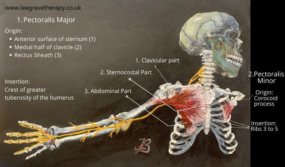
MUSCLE ANATOMY – PECTORALIS MAJOR
Click on Image to Enlarge
 1
1
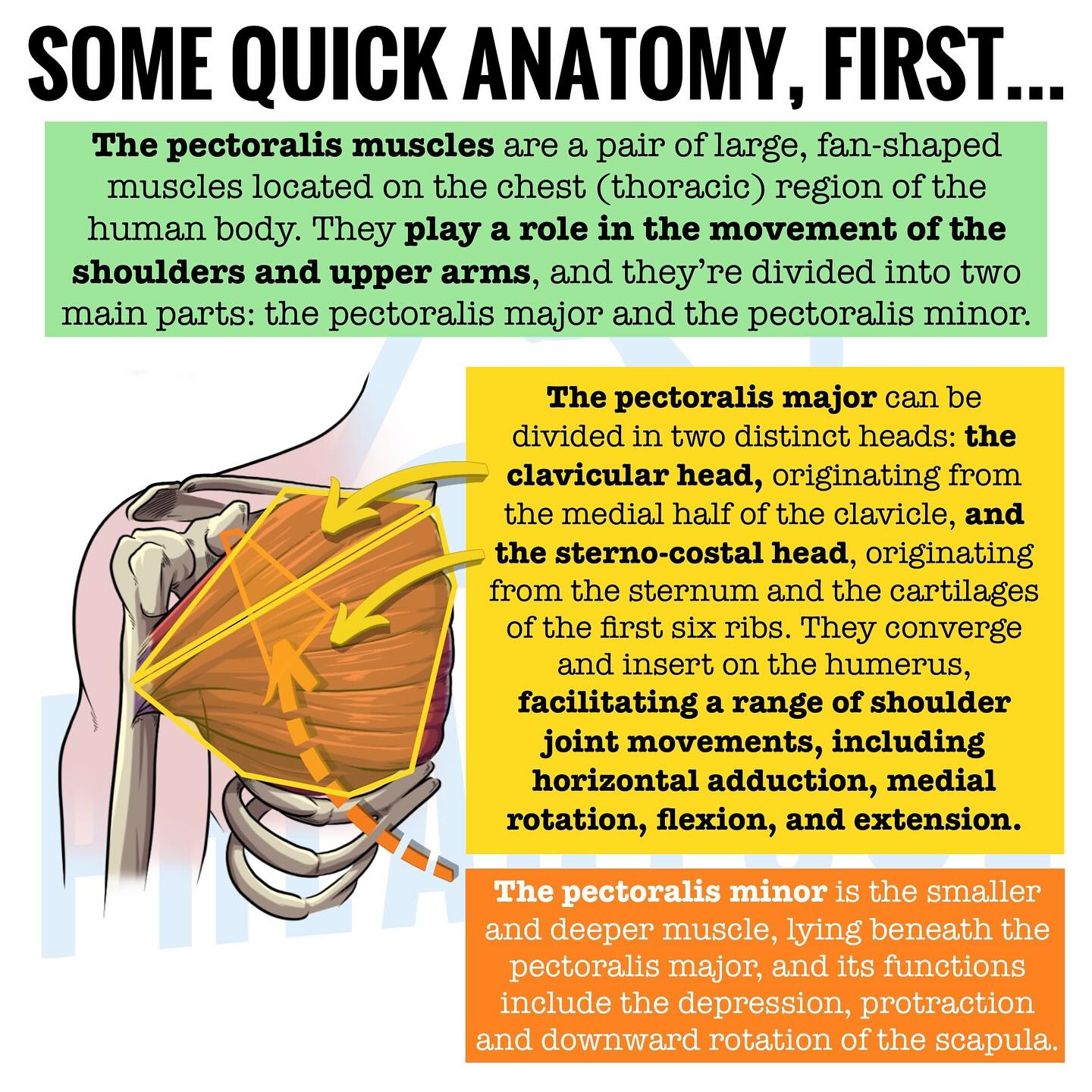
- CLAVICULAR – UPPER or SUPERIOR REGION
(1) Shoulder Flexion (2) Ab·duction above shoulder height (3) Horizontal Ad·duction (Horizontal Flexion) (4) Internal (Medial) Rotation - STERNAL – MIDDLE REGION
(1) Shoulder Ad·duction (2) Horizontal Ad·duction (Horizontal Flexion) (3) Flexion (4) Internal (Medial) Rotation - COSTAL – LOWER or INFERIOR REGION
(1) Shoulder Ad·duction (2) Horizontal Ad·duction (Horizontal Flexion) (3) Extension (4) Internal (Medial) Rotation
LINK – Normal anatomy of the pectoralis major muscle.
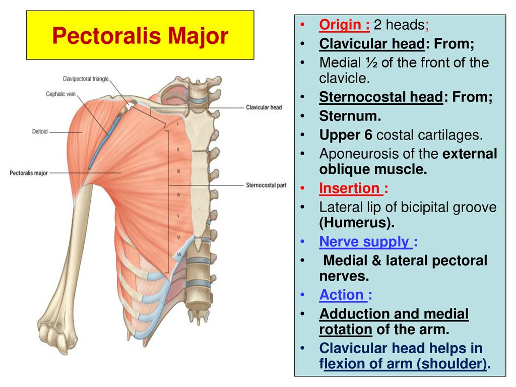
(1) Clavicular head (CH):
– the proximal clavicular head attaches to the medial half of the clavicle
(2) Sternal head – several segments (S1-S6).
– the sternal head segments attach to the sternum, second to sixth costal cartilages, and aponeurosis of the external oblique muscle.
Note: The clavicular and sternal head tendons combine to form a U-shaped tendon laterally; this tendon consists of an anterior layer (AT) and posterior layer (PT). This common tendon inserts onto the humerus at the lateral lip of the bicipital groove.
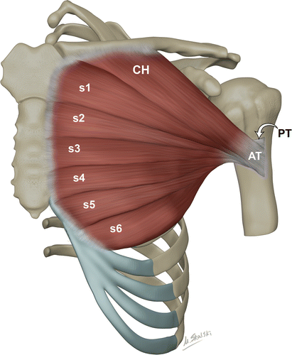
RADIO GRAPHICS .
(1) Clavicular Head (20%): is a single architectural segment that cannot be further divided. It’s within the clavicular lamina and arises from the medial half of the clavicle.
(2) Sternal head (80%): can be subdivided into 6 to 7 segments along individual fascial planes. The segments are within the abdominal and manubrial laminae and arise from the anterior manubrium, sternum, and second to sixth costal cartilages.
VIDEOS
- NOTED ANATOMIST – GLENOHUMERAL JOINT .
(1) Pectoralis Major (2) Teres Major (3) Latissimus Dorsi - ANATOMY ZONE – PECTORALIS MAJOR .
- WEBSTER – LARGE SHOULDER MUSCLES – 11:40 .